报告题目:Phase Imaging of Mechanical Properties of Live Cells活细胞力学特性的相位成像
报告时间:2018年5月22日15:00-16:30
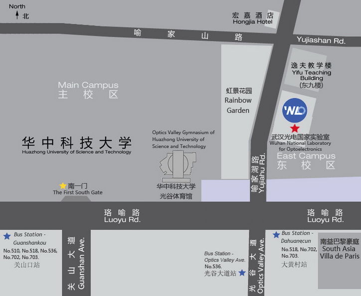
报告地点:光电国家研究中心A101
报 告 人:Prof.Adam Wax 美国杜克大学
邀 请 人:骆清铭 教授
报告人简介:
Adam Wax于1993年在位于纽约州特洛伊的伦斯勒理工学院获得电气工程学位,并且在纽约州奥尔巴尼的纽约州立大学物理系取得第二个学士学位。他分别于1996年和1999年在位于北卡罗来纳州达勒姆的杜克大学取得物理硕士和博士学位。
他曾在位于剑桥的麻省理工学院光谱实验室做博士后研究工作,现为杜克大学生物医学工程系教授和研究生院主任。他的研究兴趣包括用于早期癌症检测的光谱学,新型显微镜学和干涉测量技术。Wax博士是美国光学学会,SPIE,AIMBE的会士。他在2012年被评为“杰出博士后导师”。
Biography:
Adam Wax received the B.S. degree in electrical engineering from Rensselaer Polytecnic Institute, Troy, NY, and the second B.S. degree in physics from the State University of New York at Albany, Albany, NY, in 1993, and the M.A. and Ph.D. degrees in physics from Duke University, Durham, NC, in 1996 and 1999, respectively.
He was a Postdoctoral Fellow at George R. Harrison Spectroscopy Laboratory, Massachusetts Institute of Technology, Cambridge. He is currently Professor of Biomedical Engineering and Director of Graduate Studies at Duke University. His research interests include optical spectroscopy for early cancer detection and novel microscopy and interferometry techniques. Dr. Wax is a fellow of the Optical Society of America, SPIE and AIMBE. He was named Outstanding Postdoc Mentor in 2012.
报告摘要:
细胞对机械刺激作出反应的机制对于细胞功能研究至关重要,但尚未得到很好的理解。许多已经开发的流变学工具可以表征细胞粘度和弹性特性,但是这些工具通常需要直接的机械接触,限制了它们的通量。我们已经开发了一种新的无标记、非接触、单次成像方法,通过测量细胞的机械刚度来表征亚细胞结构的组织特征。新的分析方法可测量折射率的变化,并将其与无序强度相关联。这些测量结果与细胞刚度进行比较,使用相同的成像工具测量,以显示流动剪切刺激的纳米级响应。通过比较具有不同力学性质的五个细胞群的剪切刚度和相位无序强度来显示该技术的效用。无序强度和剪切刚度之间的相反关系,表明可以使用这种新颖的非接触技术,以适合于高通量研究的形式评估细胞机械性质。我们将进行更多的研究,其中包括检查早期致癌事件中的机械刚度和研究特定细胞结构蛋白在机械转导中的作用。
Abstract:
The mechanisms by which cells respond to mechanical stimuli are essential for cell function yet not well understood. Many rheological tools have been developed to characterize cellular viscoelastic properties but these typically require direct mechanical contact, limiting their throughput. We have developed a new approach for characterizing the organization of subcellular structures using a label free, noncontact, singleshot phase imaging method that correlates to measured cellular mechanical stiffness. The new analysis approach measures refractive index variance and relates it to disorder strength. These measurements are compared to cellular stiffness, measured using the same imaging tool to visualize nanoscale responses to flow shear stimulus. The utility of the technique is shown by comparing shear stiffness and phase disorder strength across five cellular populations with varying mechanical properties. An inverse relationship between disorder strength and shear stiffness is shown, suggesting that cell mechanical properties can be assessed in a format amenable to high throughput studies using this novel, noncontact technique. Further studies will be presented which include examination of mechanical stiffness in early carcinogenic events and investigation of the role of specific cellular structural proteins in mechanotransduction.
武汉光电论坛是武汉光电国家研究中心倾力打造的公益性高端学术系列讲座,共享光电知识盛宴,诚邀您的参与!